A Brief History of Medical Thermography

Infrared technology and Medical Thermography (recording heat variation in physiological testing) have two separate histories, but they were brought together ultimately after centuries of scientific experiments. The first person to link body temperature to physiology was Hippocrates, around 400BC, who covered patients in wet cloth and observed the different rates of drying over different regions of the body, associating the location of heat with illness and disease.
IR Detection
The idea of using light to ‘map’ temperature variation originated much later. In 1800, the astronomer Willian Hershel discovered a new spectrum of visible light, now known as infrared (IR). Over the next hundred years various ways to detect IR were devised, but recording it was far more complex. Then in 1929, the first IR sensitive camera was invented by a Hungarian physicist Kalman Tihanyi and used for anti-aircraft defences of Britain – later iterations being developed for different war-time and security uses. But much later, during the 1960s and 70s, technology paved the way for scanning devices and later image-displays on computer screens, which changed everything.
Thermographic Physiology
One of the the early medical thermography pioneers was German doctor Carl Wunderlich (1815-1877) who’s strength in physiology and methodology of diagnosis lead to his significant publication: “Temperature in Diseases, a manual of medical thermometry” in 1868. But it wasn’t until the middle of the 20th century that the first ‘Pyroscan’ instrument was used in Bath, UK to create an image of the increase in heat generated by arthritic joints. Around the same time, Dr Ray Lawson of London, Canada was the first to associate heat with breast abnormality when he discovered that breast tumours generate on average, 2 to 3 degrees F more heat than the surrounding tissue. Lawson set out to find a method to measure this heat, soon finding his inspiration from military IR technology used by heat-seeking missiles, and he devised an instrument he called the “Thermoscan”.
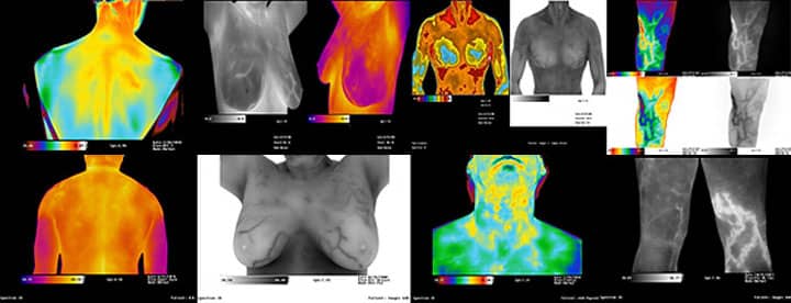
Thanks to these innovators and the modern technology of today, Thermography has become a popular diagnostic tool in Vascular Medicine, Sports Medicine, and Breast Health, capturing highly detailed images and interpreted by qualified physicians.
References
http://www.uhlen.at/thermology-international/data/pdf/223A03.pdf
https://academic.oup.com/rheumatology/article/43/6/800/1784527
https://www.ncbi.nlm.nih.gov/pmc/articles/PMC5392120/
