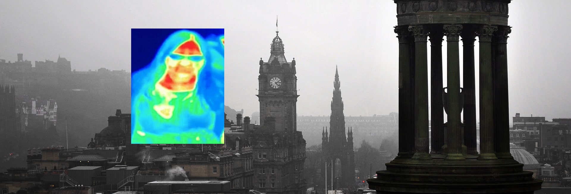
Breast Cancer Revealed by ‘Accident’ with Museum Thermal Camera

Thermal Imaging was in the news today when it was revealed that a tourist, Bal Gill, while visiting a camera museum in Scotland was captured by a thermal camera which showed one of her breasts was a different colour. On return to her home in Slough, she consulted her doctor who confirmed that she had breast cancer.
Bad Gill said: ”While making our way through the floors we got to the thermal imaging camera room. As all families do, we entered and started to wave our arms and look at the images created.
“While doing this I noticed a heat patch coming from my left breast. We thought it was odd and having looked at everyone else they didn’t have the same. I took a picture and we carried on and enjoyed the rest of the museum.”
Surprises all round, but not for us! This story confirms that Thermography works very well as a tool that indicates physiological abnormalities in the body. The BBC website reports: “Thermography, also called thermal imaging, uses a special camera to measure the temperature of the skin on the breast’s surface. It is a non-invasive test that does not involve any harmful radiation. Cancer cells grow and multiply very fast. Blood flow and metabolism are higher in a cancer tumour as blood flow and metabolism increase, which makes skin temperature rise.”
